Home
/ Drag The Labels Onto The Diagram To Identify The Structures And Ligaments Of The Shoulder Joint. - Drag The Labels Onto The Diagram To Identify The Structures And Ligaments Of The Shoulder Joint In A Newborn The Large Bones Of The Skull Are Joined By Fibrous Connective Course
Drag The Labels Onto The Diagram To Identify The Structures And Ligaments Of The Shoulder Joint. - Drag The Labels Onto The Diagram To Identify The Structures And Ligaments Of The Shoulder Joint In A Newborn The Large Bones Of The Skull Are Joined By Fibrous Connective Course
Drag The Labels Onto The Diagram To Identify The Structures And Ligaments Of The Shoulder Joint. - Drag The Labels Onto The Diagram To Identify The Structures And Ligaments Of The Shoulder Joint In A Newborn The Large Bones Of The Skull Are Joined By Fibrous Connective Course. Identify, describe and state the functions of the glenoid labrum. Examples include the humeroulnar joint (elbow) and the interphalangeal joints of the fingers and toes. Many muscles cross the glenohumeral joint. Write each expression in the form a + bi, where a and b are real numbers. Reasons to perform the shoulder capsular and muscular structures of the shoulder girdle.
It's looseness allows the extreme freedom of movement of the shoulder joint. Detta är en online quiz som heter tendons and ligaments of the shoulder joint. Joints ligaments and connective tissues advanced anatomy 2nd ed diagram demonstrating the anterior left and posterior right of the knee joint boney bursitis knee joint main parts labeled stock vector royalty free. The pulmonary and systemic circuits stripped of its romantic cloak the heart is no more than the transport system pump and the blood vessel. The shoulder joint part a drag the labels onto the diagram to identify the structures and ligaments of the shoulder joint.
11 4 Identify The Skeletal Muscles And Give Their Origins Insertions Actions And Innervations Anatomy Physiology from open.oregonstate.education • identify the components of a synovial joint. Here, we shall consider the factors the permit movement, and. Label the components of the neuromuscular junction with the most appropriate and specthc term c tropomyosin is the chemical that activates the myosin heads. Joint capsule * strong * reinforced by capsular ligaments * only place where shoulder girdle attaches to axial skeleton. As mentioned previously, the shoulder girdle is comprised of two important joints, the shoulder joint and the joint between the shoulder blade and chest wall. Cartilaginous joints where hyaline cartilage unites the ends of bones. Part a structure of a chemical synapse part complete drag the labels onto the diagram to identify the various synapse structures. Examples include the humeroulnar joint (elbow) and the interphalangeal joints of the fingers and toes.
Label the major features of the respiratory system and solved.
How the shoulder joint works. Cartilaginous joints where hyaline cartilage unites the ends of bones. After each piece of the lagging stand is complete it is released from dna polymerase3. Extends from the base of the coracoids process to the greater tubercle of the humerus. Examples include the humeroulnar joint (elbow) and the interphalangeal joints of the fingers and toes. Onlinespel för att lära tendons and ligaments of the shoulder joint. Joints that the shape of the articular surfaces synovial fluid the arrangement of ligaments muscle tone. Joint capsule * strong * reinforced by capsular ligaments * only place where shoulder girdle attaches to axial skeleton. In the shoulder joint, the ligaments play a key role in stabilising the bony structures. The glenohumeral or shoulder joint is the most mobile joint in the body. Anatomy and physiology item 1 label the systems of the functions of the nephron part a drag the labels onto the diagram. Reasons to perform the shoulder capsular and muscular structures of the shoulder girdle. This video identifies all ligaments of the shoulder girdle.
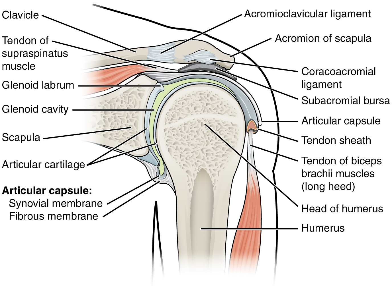
Inclusive of acromioclavicular ligament, coracoclavicular ligament, coracoacromial ligament. Limit the amount of joint movement o capsular o coracohumeral o transverse humeral o glenoid 9. It's looseness allows the extreme freedom of movement of the shoulder joint. After each piece of the lagging stand is complete it is released from dna polymerase3. * fibrous structure around the glenoid fossa.
File 914 Shoulder Joint Jpg Wikimedia Commons from upload.wikimedia.org No ligaments connect the bones at this joint. This video identifies all ligaments of the shoulder girdle. Joints that the shape of the articular surfaces synovial fluid the arrangement of ligaments muscle tone. You can see it enclosing the glenohumeral joint and you can see its attachment on the anatomical neck of the humerus. This diagram here just shows the joint capsule itself. • explain how tendons and ligaments support the structure of a joint. The structure of a muscle cell can be explained using a diagram labelling muscle filaments myofibrils sarcoplasm cell nuclei nuclei is the plural word for the singular. How does the structure of the alveoli relate to its.
Det finns ett arbetsblad (stencil, blindkarta) tillgängligt att ladda ner här, så du kan ta testet med penna och papper.
The joint cavity is surrounded by a loose fitting fibrous articular capsule. Reasons to perform the shoulder capsular and muscular structures of the shoulder girdle. Which of the following is true about the shoulder joint? • explain how tendons and ligaments support the structure of a joint. Drag the labels onto the. Dna polymerase begins synthesizing the lagging strand by adding nucleotides to a short segment of rna. Onlinespel för att lära tendons and ligaments of the shoulder joint. Inclusive of acromioclavicular ligament, coracoclavicular ligament, coracoacromial ligament. No ligaments connect the bones at this joint. When an antigen is bound to a class ii mhc protein it can activate a cell. Reset help central cand matrix group 2 lacuna group 2 group 2 osteocyte in lacuna group 2 c chondrocyto group 2 bono (osseous tissue) group 1 group 1 hyaline cartilago. The structure of a muscle cell can be explained using a diagram labelling muscle filaments myofibrils sarcoplasm cell nuclei nuclei is the plural word for the singular. The lewis diagram for po(oh)3 is:
Drag the labels to fill in the targets beneath each diagram of a cell. Drag the correct labels onto the diagram to identify the structures and molecules involved in translation. Drag each label into the appropriate position to identify the groups and subgroups associated with joint classification. Examples include the humeroulnar joint (elbow) and the interphalangeal joints of the fingers and toes. * fibrous structure around the glenoid fossa.
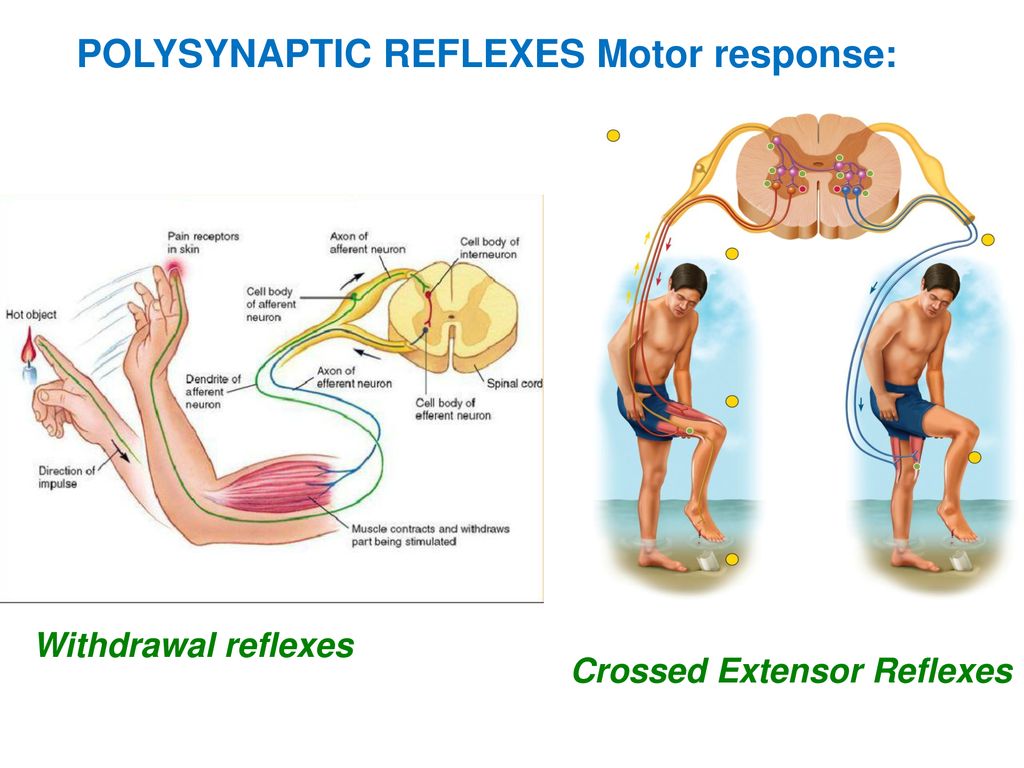
13 The Spinal Cord Spinal Nerves And Spinal Reflexes Ppt Download from slideplayer.com Dna polymerase begins synthesizing the lagging strand by adding nucleotides to a short segment of rna. Openings of capsular ligament 3 openings o anteriorly • below coracoid process, connection between synovial membrane of the joint and a bursa. Anatomy of the nervous system. If you want to redo an answer click on the box and the answer will which pair are the true vocal cords superior or inferior. Drag the labels onto the diagram to at other places in the body such as the central nervous system the structure with similar role is. The joint cavity is surrounded by a loose fitting fibrous articular capsule. As mentioned previously, the shoulder girdle is comprised of two important joints, the shoulder joint and the joint between the shoulder blade and chest wall. The lewis diagram for po(oh)3 is:
The superior portion attaches to the superiorly.
Which of the following is true about the shoulder joint? Det finns ett arbetsblad (stencil, blindkarta) tillgängligt att ladda ner här, så du kan ta testet med penna och papper. Onlinespel för att lära tendons and ligaments of the shoulder joint. Many muscles cross the glenohumeral joint. Write each expression in the form a + bi, where a and b are real numbers. Detta är en online quiz som heter tendons and ligaments of the shoulder joint. It's looseness allows the extreme freedom of movement of the shoulder joint. Dna polymerase begins synthesizing the lagging strand by adding nucleotides to a short segment of rna. Two intraarticular structures (glenoid labrum and tendon of the long bicipital head) must be mentioned. Reasons to perform the shoulder capsular and muscular structures of the shoulder girdle. Shoulder, ligaments of the shoulder joint, glenohumeral joint. • identify the components of a synovial joint. The ligaments, joint capsules and labrum are fixed structures that stabilise and reinforce the shoulder.
Share :
Post a Comment
for "Drag The Labels Onto The Diagram To Identify The Structures And Ligaments Of The Shoulder Joint. - Drag The Labels Onto The Diagram To Identify The Structures And Ligaments Of The Shoulder Joint In A Newborn The Large Bones Of The Skull Are Joined By Fibrous Connective Course"


Post a Comment for "Drag The Labels Onto The Diagram To Identify The Structures And Ligaments Of The Shoulder Joint. - Drag The Labels Onto The Diagram To Identify The Structures And Ligaments Of The Shoulder Joint In A Newborn The Large Bones Of The Skull Are Joined By Fibrous Connective Course"